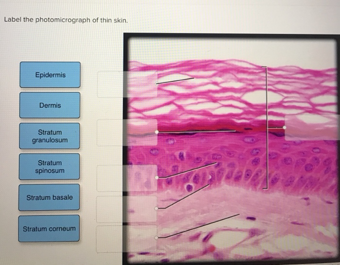
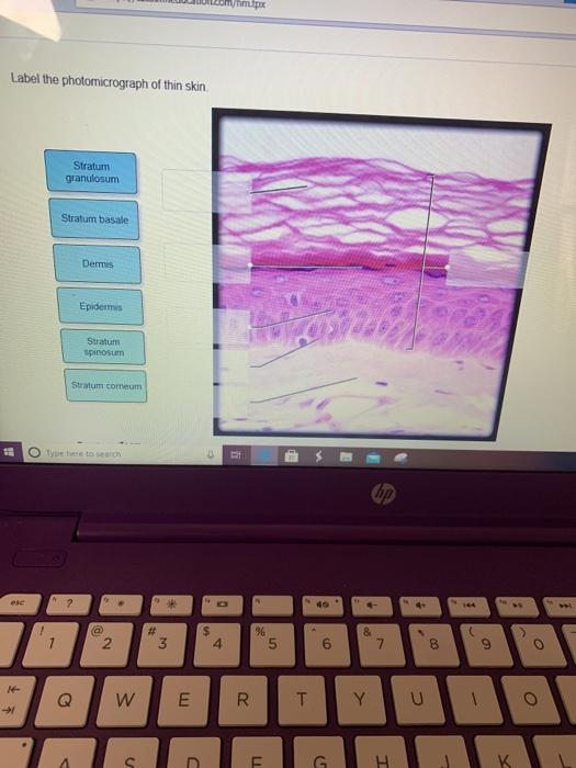
39 label the photomicrograph of thin skin.
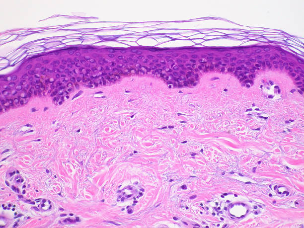
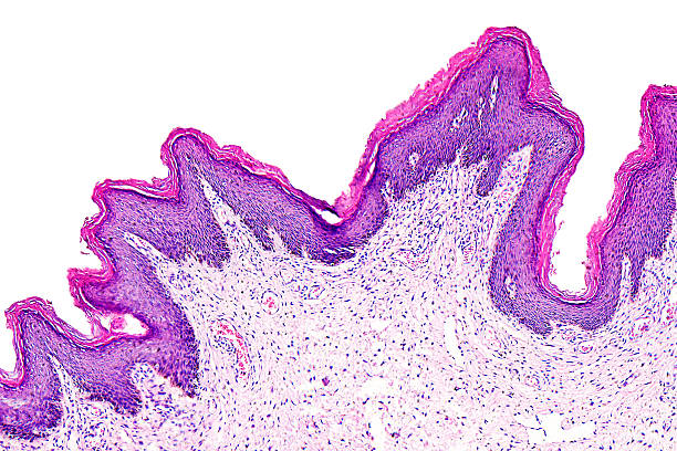
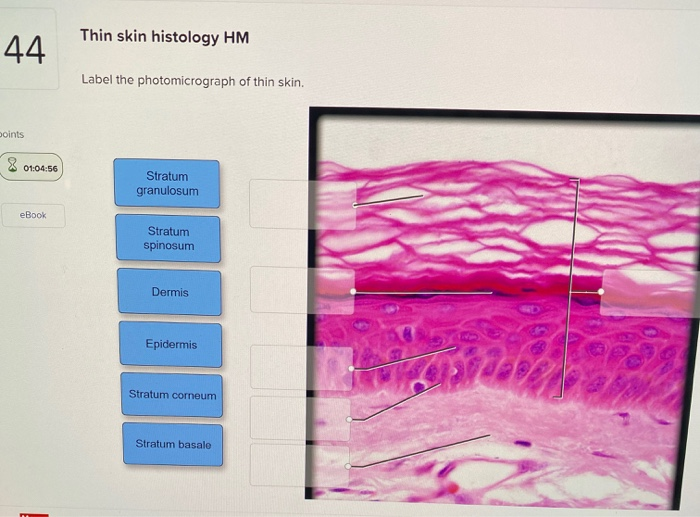
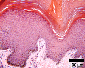
Skin overview 4 | Digital Histology Thin skin. Skin can be classified as either thick or thin, depending on the thickness of the epidermal layer. A diagrammatic representation of thin skin and a photomicrograph of a H&E stained section illustrate the reduced thickness of the epidermal strata in thin skin and the absence of stratum lucidum as a distinct layer. 400x. anatomy lab, exam 3, lab 9, Spinal Nerves, Integument, and ... - Quizlet Label the photomicrograph of thin skin. stratum corneum stratum granulosum stratum spinosum stratum basale dermis epidermis hypodermis the layer of skin beneath the dermis, which serves as a storage repository for fat Name the yellow highlighted structures that pass through the intervertebral foramina. spinal nerves
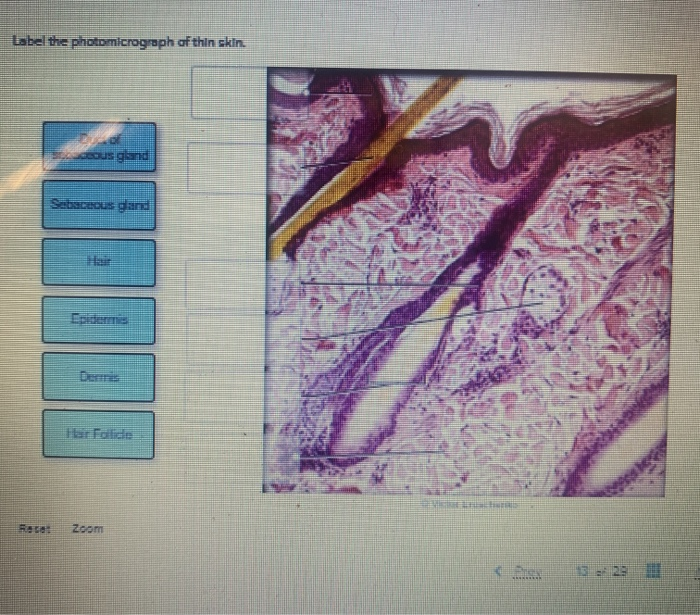
photomicrographs of thin skin Flashcards | Quizlet photomicrographs of thin skin stratum corneum Click the card to flip 👆 Click the card to flip 👆 1 / 4 Flashcards Learn Test Match Created by Madison_Tacquard Terms in this set (4) stratum corneum sebaceous gland hair follicle dense irregular CT of the reticular layer of the dermis Students also viewed LP5 LAB 6 terms jordanmmertens

Label the photomicrograph of thin skin.
(Solved) - Label the photomicrograph of thin skin. Label the ... Label the photomicrograph of thin skin 1 Approved Answer Mohinee k answered on August 05, 2021 4 Ratings ( 9 Votes) Label the photomicrograph of the skin A photograph taken with the help of microscope . Skin is the largest sensory organ in body.its protect the body sense pain... solution .pdf Label the photomicrograph of thin skin. O Stratum granulosum O Stratum ... Label the photomicrograph of thin skin. Stratum granulosum Stratum spinosum Dermis Epidermis Stratum corneum Stratum basale We store cookies data for a seamless user experience. Layers of the Skin | Anatomy and Physiology I - Lumen Learning Most of the skin can be classified as thin skin. "Thick skin" is found only on the palms of the hands and the soles of the feet. It has a fifth layer, called the stratum lucidum, located between the stratum corneum and the stratum granulosum (Figure 2). Figure 2. Thin Skin versus Thick Skin.
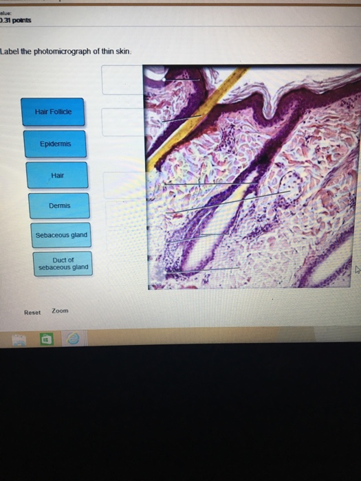
Label the photomicrograph of thin skin.. Label the photomicrograph of thin skin. Dermis Duct of ... - Brainly The skin is the largest organ in the body; it extends from the inside of the body to the outside, has a thickness of about two millimeters, and weighs around six pounds on average. It protects the body against excessive heat and light, as well as injury and infection. Layers of the Skin | Anatomy and Physiology I - Lumen Learning Most of the skin can be classified as thin skin. "Thick skin" is found only on the palms of the hands and the soles of the feet. It has a fifth layer, called the stratum lucidum, located between the stratum corneum and the stratum granulosum (Figure 2). Figure 2. Thin Skin versus Thick Skin. Label the photomicrograph of thin skin. O Stratum granulosum O Stratum ... Label the photomicrograph of thin skin. Stratum granulosum Stratum spinosum Dermis Epidermis Stratum corneum Stratum basale We store cookies data for a seamless user experience. (Solved) - Label the photomicrograph of thin skin. Label the ... Label the photomicrograph of thin skin 1 Approved Answer Mohinee k answered on August 05, 2021 4 Ratings ( 9 Votes) Label the photomicrograph of the skin A photograph taken with the help of microscope . Skin is the largest sensory organ in body.its protect the body sense pain... solution .pdf
BSC2085 - Ch. 05 Module 1 Section 5 01-5.02 Dynamic Study ...
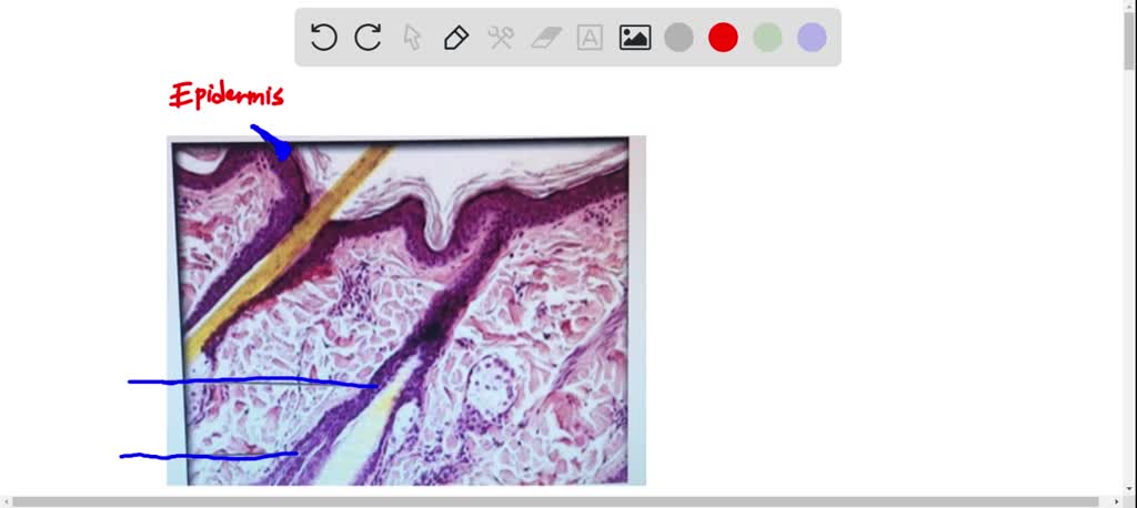
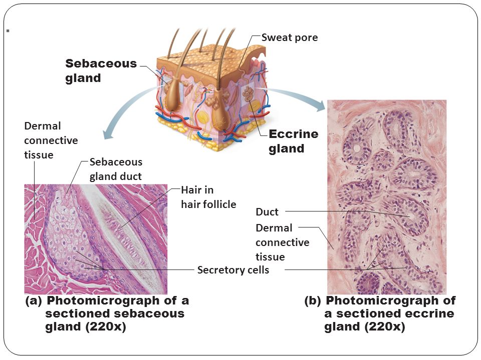
Label tne photomicrograph Of the Skin and Its accessory structures, Sebaceous gland, Duct ofl, sebaceous gland, Epidermis, Hair follicle
Solved Label the photomicrograph of thin skin. Epidermis ...

BIOL 319 Lab 1 Flashcards | Quizlet

A rare case report of apixaban-induced lichenoid eruption ...
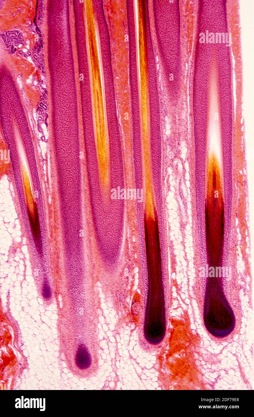
Hair follicle micrograph hi-res stock photography and images ...

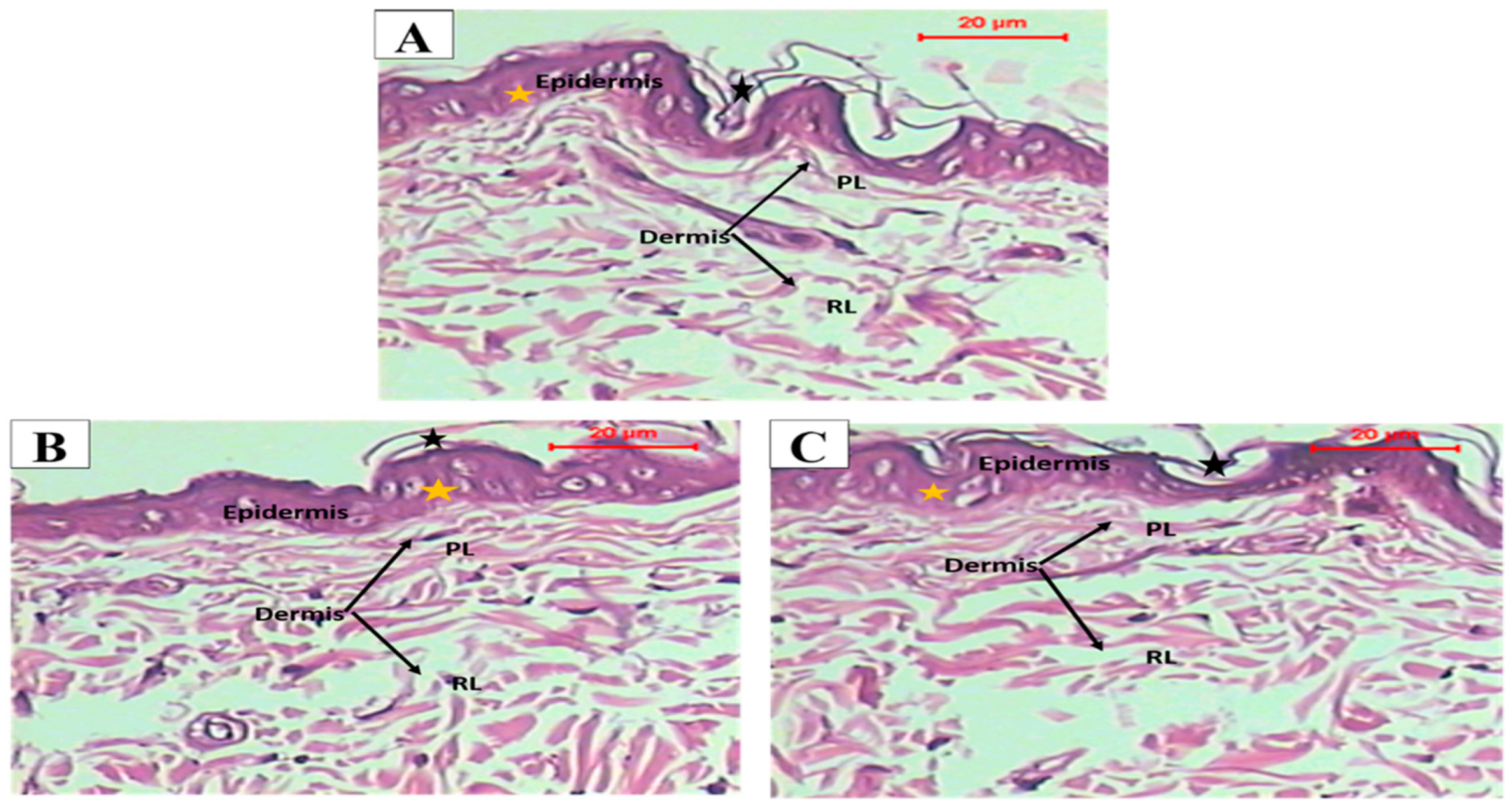
Photomicrographs of thin skin sections. (A) A section in thin ...
2,100+ Skin Histology Stock Photos, Pictures & Royalty-Free ...
Skin and the Integumentary System

View Image
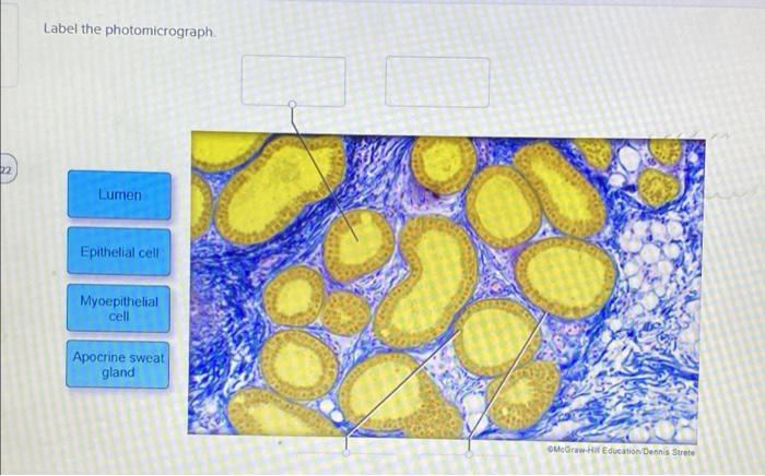
Solved Label the photomicrograph. Lumen Epithelial cell ...

a) A photomicrograph of the section of thin skin tissue from ...
Integumentary System Overview
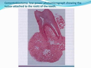
Odontegenic tumors
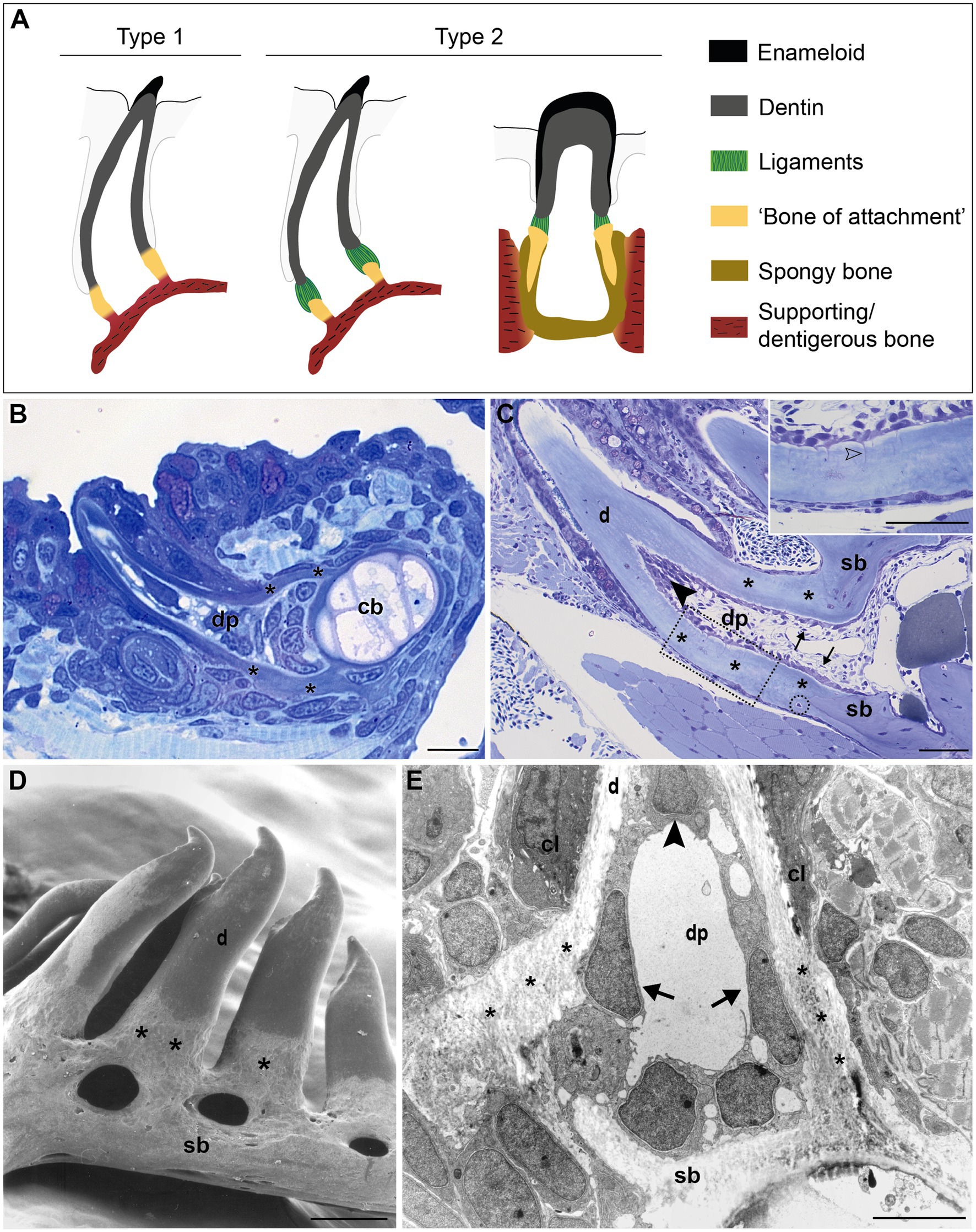
Frontiers | Cells at the Edge: The Dentin–Bone Interface in ...

Figure 7.4 Photomicrograph of the skin and accessory ...
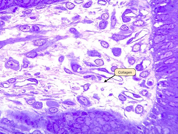
Connective Tissue Lab

Figure 5 | Time-Dependent Effect of Oral Morphine Consumption ...

BIOL 319 Lab 1 Flashcards | Quizlet
2,100+ Skin Histology Stock Photos, Pictures & Royalty-Free ...
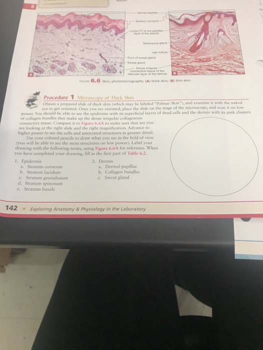
6.6 Skin, photomicrographs: tA) thick skin; OB) thin | Chegg.com
Pharmaceutics | Free Full-Text | Development of Transdermal ...
Animals | Free Full-Text | Naturally Produced Lovastatin ...
Solved met Label the photomicrograph of thin skin Stratum ...
Light photomicrograph hi-res stock photography and images - Alamy
Solved Label the photomicrograph of thin skin. | Chegg.com
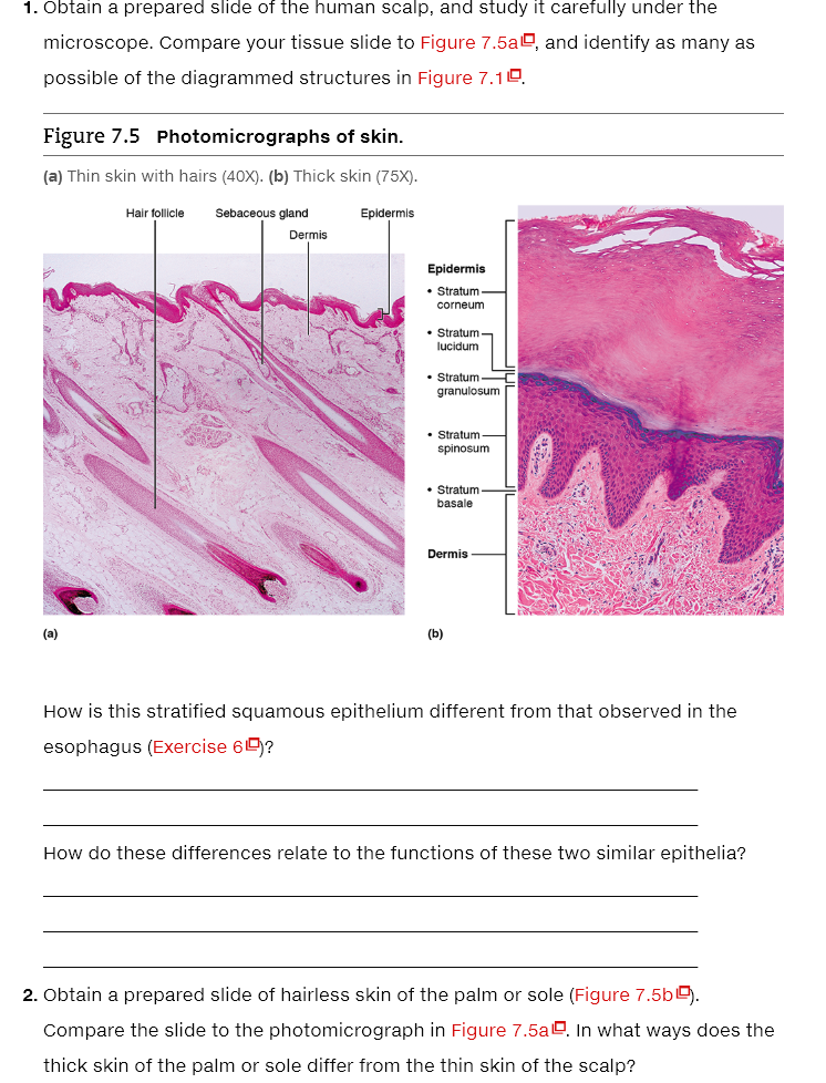
Solved 1. Obtain a prepared slide of the human scalp, and ...

Pin by nico x. on Anatomy | Games, Tetris, Anatomy

Lab 9: Pre-Lab Homework Flashcards | Quizlet

View Image
Solved Thin skin histology HM 44 Label the photomicrograph ...
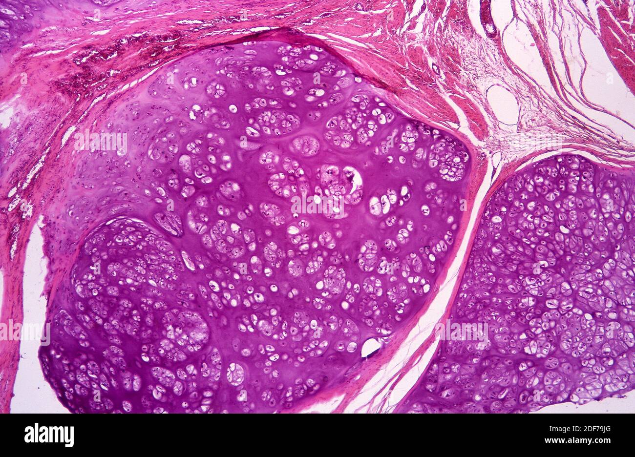
Cartilaginous tumor hi-res stock photography and images - Alamy
Hair shaft Dermal papillae Epidermis Subpapillary vascular ...
Solved Label the photomicrograph of thin skin. deous gland ...
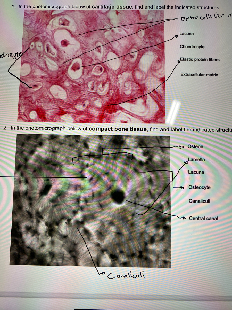
Answered: 1. In the photomicrograph below of… | bartleby

Epidermis | Biology for Majors II
Solved] Label The structures of the bone. Femur Lateral ...
Skin: The Histology Guide

Blood
Post a Comment for "39 label the photomicrograph of thin skin."